2026-09-26
What If the Chest Depresses After Gynecomastia Surgery? Understanding Chest Indentation Complications
Explains why preserving 5–10 mm of subareolar tissue during gynecomastia surgery is critical to prevent chest indentation (saucer deformity) and maintain a natural contour.
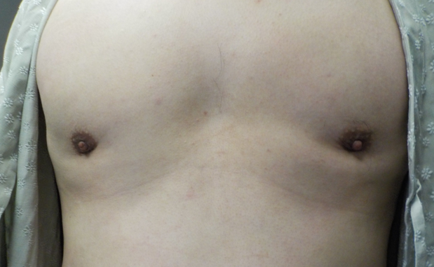
Hello, I am Dr. Seong-il Cho, a general surgeon.
When consulting patients about gynecomastia surgery, I encounter two questions most frequently that seem entirely contradictory.
"Doctor, will all the glandular tissue be completely removed during surgery?"
VS
"I heard the chest can indent after surgery. Is it really safe to remove all the glandular tissue?"
Considering the long-term emotional distress patients suffer from protruding chest contours, I fully understand the desire to eradicate every bit of the problematic glandular tissue without leaving a trace. Simultaneously, fears of complications such as localized chest depression after surgery are just as real, leading to many anxious inquiries.
To summarize before delving into the details: completely removing 100% of the glandular tissue down to the muscle floor can cause serious cosmetic side effects and must be avoided. The standard, appropriate approach—employed in the vast majority of operations—is to leave a normal, physiologic baseline amount of glandular tissue.
Let us explore this step by step. This article addresses why a certain volume of glandular tissue must be preserved during gynecomastia surgery, along with the surgical techniques involved.
1. What Happens When Glandular Tissue Is Completely Eradicated: Saucer Deformity
Beneath the nipple and areolar complex lies an inherently thin layer of fat and glandular tissue. These tissues are not merely targets for excision; they function as a foundational support structure preventing the nipple and areola from sinking inward.
What happens if the subareolar glandular tissue is aggressively scooped out down to the very base purely to prevent recurrence?
While the chest might appear flat immediately post-op, healing leads to scar contracture and the skin adhering directly to the underlying pectoralis major muscle, causing the nipple and areolar complex to sink and hollow out drastically.
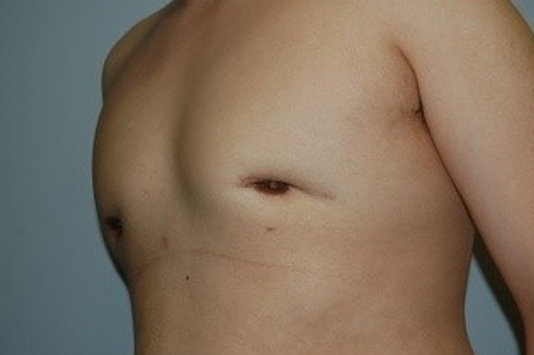
In medical terminology, this is known as a 'saucer deformity' or 'nipple-areolar depression.' Rather than achieving a smooth, natural chest contour, the peri-areolar area indents like the bottom of a pan or saucer, often causing far more aesthetic distress than the original condition before surgery.
2. The Medically Recommended Standard for Glandular Preservation
Then, precisely how much glandular tissue is surgically safest and most ideal to preserve?
According to standard plastic and general surgery reference guidelines (Plastic Surgery Key - Gynecomastia Surgical Techniques), the principle is defined as follows:
"When removing the subareolar mass by direct excision, leaving enough tissue (approximately 5 to 10 mm of evenly layered tissue) under the nipple is critical to prevent a central depression or saucer deformity."
In standard surgical practice, an even subareolar disc of glandular and soft tissue measuring approximately 5 to 10 mm must be preserved directly beneath the areola.
This 5 to 10 mm layer acts as a critical cushion to uphold the structural integrity of the nipple, ensuring that the chest profile remains flat and natural without post-surgical hollowing.
3. Could Preserving Glandular Tissue Cause Recurrence?
You might wonder: "If 5 to 10 mm is left behind, won't gynecomastia simply recur later from that remaining tissue?"
To answer directly, the 5 to 10 mm tissue preserved during surgery is not a pathological mass, but physiological tissue required to maintain normal anatomical structure.
Gynecomastia does not recur simply because minimal glandular tissue remains. Unless rare triggers intervene—such as long-term use of specific medications inducing severe hormonal imbalances or extreme weight fluctuations—the likelihood that this preserved tissue will hypertrophy again into enlarged breasts is exceptionally low.
Conversely, correction of chest depression caused by over-resection is notoriously complex, often requiring secondary fat grafting or intricate local flap procedures. Therefore, the surgical precision to retain the correct volume of glandular tissue during the primary surgery is paramount.

4. Surgical Expertise: Beyond Resection to Contour Reconstruction
Preserving a uniform 5 to 10 mm layer of tissue is far from simple in clinical practice. Skin thickness, adipose volume, and glandular tissue density vary widely across individual patients.
A seasoned surgeon continuously conducts pinch tests to evaluate flap thickness throughout the procedure, simultaneously employing feathering techniques to eliminate abrupt steps between the preserved subareolar tissue and surrounding liposuction margins.
Gynecomastia surgery is not simply about carving out internal tissue. It is a precise reconstructive procedure designed to yield a flat, smooth, three-dimensional masculine contour tailored to the patient's overall chest proportions and skin thickness.
Rather than evaluating a procedure quantitatively by asking "How much gland was excised?" or "How much fat was suctioned?", the crucial factor is "How harmoniously was the excision balanced to match my individual chest shape?"
This article addresses the two opposing questions: "What if the areola indents?" and "What if it still bulges after surgery?"
While these are common concerns for patients, the operating surgeon understands these subtleties deeply—often agonizing over and planning these precise contours even more thoroughly than the patients themselves.
Thank you for taking the time to read this.
References
Gynecomastia: Surgical Management and Technical Considerations (Plastic Surgery Key / Educational Guidelines) [Link: https://plasticsurgerykey.com/gynecomastia-4/]
Correction of areolar depression in postsurgically treated gynecomastic patients (Annals of Plastic Surgery, PMID: 10213411) [Link: https://pubmed.ncbi.nlm.nih.gov/10213411/]
Phone: 031-696-1212
Gynecomastia Surgery Hotline (Call/SMS): 010-3493-0013
KakaoTalk ID: theu114
KakaoTalk Channel: Bundang The U
Frequently Asked Questions
Should glandular tissue be completely excised during gynecomastia surgery?
No, it should not be removed completely. Eliminating 100% of the tissue removes the structural foundation supporting the nipple and areola, causing the skin to adhere directly to the muscle and leading to a saucer deformity.
Why does the saucer deformity complication occur?
It occurs when subareolar glandular tissue is over-resected down to the muscle bed. As tissue heals, scarring and contraction cause the skin to bind to the pectoralis major, indenting the peri-areolar region like a plate or basin.
How much glandular tissue is safely preserved during gynecomastia surgery?
Leaving an even tissue layer of approximately 5 to 10 mm beneath the nipple is recommended. This thin tissue layer functions as a necessary cushion to support nipple projection and achieve a natural, smooth chest contour.
Will gynecomastia recur because of the remaining glandular tissue?
The likelihood of recurrence is extremely low. The 5 to 10 mm preserved is non-pathologic tissue maintained for anatomical support, which will not regrow into enlarged breasts absent extreme hormonal disturbances or severe weight changes.
Can chest depression after surgery be easily corrected?
Correction is technically demanding. Secondary revision for over-resection often necessitates fat grafting or complex flap procedures, underscoring the critical importance of proper glandular preservation during the primary procedure.